
Due to the location of the spinal nerves, vertebrae, and discs deep within the body, any strategy to obtain access to the spinal area necessitates the removal of muscle tissue. This is generally accomplished by making small incisions and guiding equipment and/or microscopic video cameras through them. Lasers are rarely employed in MIS operations, contrary to popular assumption.
During MIS surgery, a variety of techniques can be employed to reduce trauma. Following are some of the more frequent techniques explained by the best spine surgeon in Mumbai.
Using a Tubular Retractor
Rather than cutting directly through the muscles, this approach includes gradual dilatation of the soft tissues. The surgeon can work through the incision without having to expose the area too much by utilizing tubes to keep the muscles out of the way. An endoscope or microscope directed down the tube may be utilized by the neurosurgeon to aid in executing the surgery using a limited access technique. The tubular retractor can be removed when the treatment is finished, allowing the dilated tissues to heal. Incisions can vary in size depending on the extent and type of surgery required.
Percutaneous Placement of Screws and Rods
Depending on the patient’s condition, instrumentation such as rods and screws may be used to support the spine or immobilize the spine to aid in the fusing of the spinal bones. Traditional screw placement methods necessitate substantial muscle and other tissue removal from the spine’s surface.
Percutaneous (meaning “through the skin”) implantation, on the other hand, usually entails introducing rods and screws through small incisions in the skin without damaging or dissecting the underlying muscle. Guidewires are inserted through the skin and into the spinal vertebrae along the intended screw pathways using x-ray pictures. The guidewires are then covered with screws that follow the wires’ course. These screws feature temporary extenders that extend outside of the skin and are removed after assisting in the connection and securement of the screws by guiding the passage of rods. Spinal instrumentation is being implanted more safely and accurately because of the use of spinal navigation and robots.
Direct Lateral Access Routes
Because there is less muscular tissue blocking the way in some cases, especially those involving the lumbar spine, accessing the spine from the side of the body can result in less pain. The patient is usually on his or her side throughout this procedure. Then, on the side of the spine, a tubular retractor docks to allow access to the spine’s discs and bones.
The Thoracoscopic Access Route
It may be essential to reach the front sections of the thoracic spine, which are positioned in the chest and are surrounded by the heart and lungs, depending on the patient’s condition. Traditional access methods frequently entail making major incisions in the chest, which may necessitate the removal of one or more ribs. Thoracoscopic access, on the other hand, relies on a series of small incisions through which functioning ports and cameras can be introduced to aid operation.
Treatment Options for MIS Surgery
For MIS surgery, a number of particular approaches have been used. Despite the fact that the area is still evolving, the list below illustrates some of the most frequent alternatives.
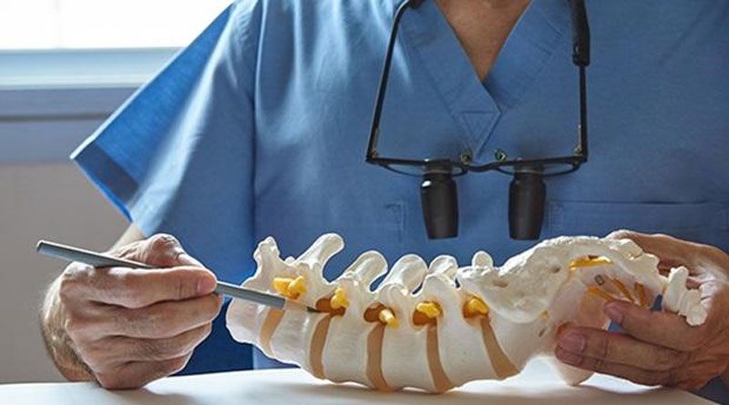
- Discectomy: Spinal discs are elastic bands containing soft material that act as cushions between the vertebral bones. The soft tissue inside the elastic ring might protrude — or herniate — outside of the ring if it becomes weaker. The debris from a herniated disc might compress nearby nerves, causing pain. If surgery to trim or remove the herniated disc is recommended, it may be possible to do so with MIS surgery employing tubular dilators and a microscope or endoscope.
- Decompression of the spine: Spinal stenosis, or a narrowing of the spinal canal, is a frequent disorder that causes nerve compression. Pain, numbness, and muscle weakness are some of the symptoms that can result. If surgery is necessary, a MIS technique using tubular dilators and a microscope or endoscope may be used to remove the bone and soft tissues that are causing the nerve compression. Laminectomy and foraminotomy are two of the most popular decompressive surgeries.
- TLIF (Transforaminal Lumbar Interbody Fusion): It is a minimally invasive procedure used to treat patients with intractable mechanical low back and radicular pain caused by spondylolisthesis, degenerative disc disease, or recurrent disc herniation. With the patient on his or her stomach, the surgery is conducted from the back (posterior). Screws and rods are put between two or more spinal levels using two tiny incisions. The intervertebral disc is removed, and a bone cage is inserted in its place, with the purpose of stabilizing the afflicted levels.